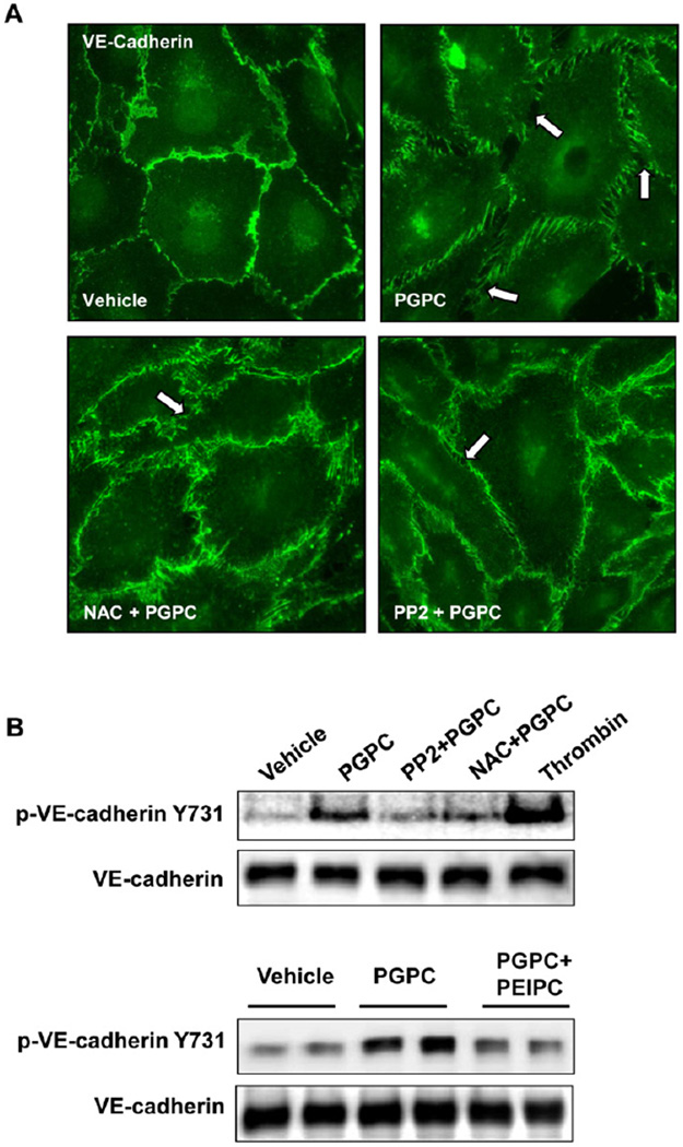
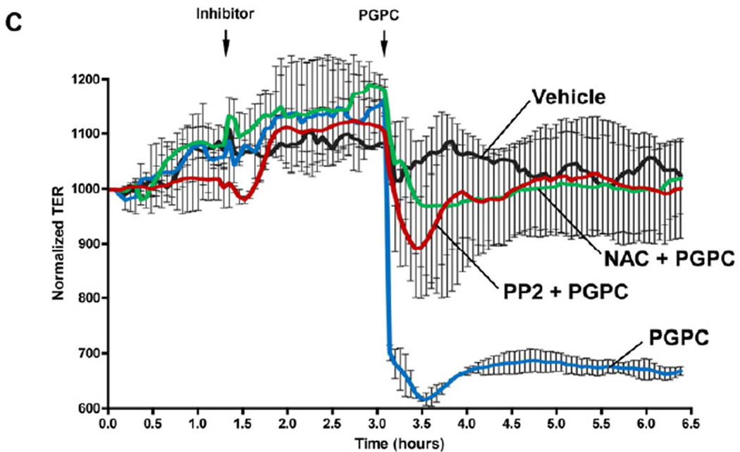
Figure 5. Effects of SRC and ROS inhibitors on remodeling of VE-cadherin-positive adherens junctions, VE-cadherin phosphorylation and EC permeability induced by PGPC.
HPAECs were pretreated with vehicle, PP2 (1µM), or NAC (1mM) for 30 min followed by stimulation with 30 µg/ml PGPC. A – Immunofluorescence staining for VE-cadherin was performed after 15 min of PGPC stimulation. Arrows depict areas of intercellular gaps. Shown are representative results of three independent experiments. B – The levels of VE-cadherin phosphorylated at Tyr731 and Y658 were detected by Western blot analysis and normalized to the total VE-cadherin content. Lower panels depict effect of co-treatment with PEIPC (2 µg/ml) on PGPC-induced VE-cadherin phosphorylation at Y731. C – TER measurements were monitored over 6.5 hrs. Time points of inhibitor and PGPC addition are indicated by vertical arrows. Shown are pooled data from three independent experiments expressed as mean ± SD.


