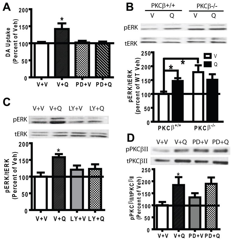
Fig. 5.
ERK activation by quinpirole required PKCβ. (A) A 15-min pretreatment with PD98059 (PD, 10 μM) blocked quinpirole (Q, 1 μM, 5 min)-stimulated DA uptake in striatal synaptosomes from PKCβ+/+ mice compared to vehicle (V) treatment. (B) Percoll-purified striatal synaptosomes from PKCβ+/+ and PKCβ−/− mice were treated with V or 10 μM quinpirole (Q) for 5 min (N=5). The upper panel shows representative blots of pERK (phosphorylated ERK) and tERK (total ERK) from a representative PKCβ+/+ and PKCβ−/− mouse. Data were converted as a ratio of pERK to tERK as a measurement of ERK activity, and calculated as a percent of the V treatment in PKCβ+/+ mice. Quinpirole significantly increased the pERK level in PKCβ+/+ but not in PKCβ−/− mice. The basal pERK level was significant higher in PKCβ−/− mice as compared to that from PKCβ+/+ mice (p<0.05). (C) Percoll-purified striatal synaptosomes from PKCβ+/+ mice were pretreated with the PKCβ inhibitor LY379196 (LY, 100 nM) or vehicle (V) for 1 hr, and the level of pERK in response to quinpirole (Q, 1 μM, 5 min) was determined (N=8). Representative blots of pERK and tERK were shown. LY379196 did not change the basal pERK, and blocked quinpirole-stimulated pERK. (D) Percoll-purified striatal synaptosomes were pretreated with the ERK inhibitor PD98059 (PD, 10 μM, 15 min) before vehicle or quinpirole treatment (Q, 1 μM, 5 min). The activity of PKCβII was determined by pPKCβII (phosphorylated PKCβII) vs. tPKCβII (total PKCβII) (N=8). Representative blots of pPKCβII and tPKCβII blots are shown in the upper panel. PD98059 itself did not change the basal pPKCβII level and did not block quinpirole-stimulated pPKCβII. Error bars represent SEM.

