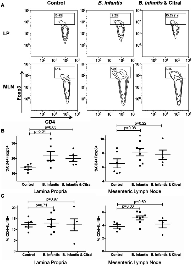
Figure 4. B. infantis induction of Foxp3+ lymphocytes is not RALDH-dependent.
(a) Representative flow cytometric dot-plots are illustrated for CD4 and Foxp3 populations within MLN and LP. (b) The increase in CD4+Foxp3+ T lymphocytes following B. infantis feeding in the LP (n = 6) is not reversed by citral treatment (n = 5). (c) CD4+IL-10+ T lymphocytes increased within MLN (n = 8), but not the LP (n = 7). LP statistical significance was estimated using the non-parametric Mann-Whitney test, while MLN statistics were determined using the parametric unpaired student t-test.

