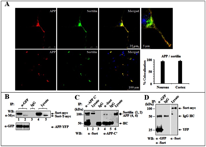
Figure 1. Sortilin and APP are allied in vitro and in vivo.
(A) Colocalization of sortilin and APP. Mouse cortical neuron (upper) and brain cortex (lower) were immunostained for APP (red) and sortilin (green). Colocalization of sortilin and APP was indicated in merged panels (yellow). DAPI stained cell nuclei (blue). Plotted colocalization: 92% mean± SEM, n = 20, in cortical neurons and 95% mean± SEM, n = 3, in brain cortexes. Scale bar 25 µm for cortical neurons, 5 µm for enlarged image and 100 µm for brain cortex. (B) Co-IP of sortilin with APP in co-transfected HEK293 cells. HEK293 cells growing in 10 cm culture dishes were co-transfected with APP770-YFP/Sort-FL-myc/His (lane 1, 4) or Sort-T-myc/His (lane 2, 5). Cell lysates were immunoprecipitated with rabbit anti-GFP (α-GFP) for APP and blotted with mouse anti-Myc (α-Myc) for sortilin. Mixed lysates were used for IgG (lane 3). Sort-FL-myc.His (Sort-myc), Sort-T-myc.His (sort-T-myc) and APP770-YFP (APP-YFP) are indicated by arrows. (C) Co-IP of sortilin with APP in APPSwe/PS1dE9 transgenic mouse brain lysate. Mouse brain lysates were subjected to immunoprecipitation with rabbit anti-APP C’ (α-APP C’) and blotted with rabbit anti-sortilin (α-Sort) (left panel) or immunoprecipitation with α-Sort and blotted with α-APP C’ (right panel). Sortilin and APP are indicated by arrows. (D) Control for Co-IP using pEYFP. HEK293 cells were co-transfected with pEYFP/Sort-myc. Co-IP was performed using α-GFP and blotted with α-sort and α-GFP. Rabbit IgG (IgG) was used as a control for non-specific binding.

