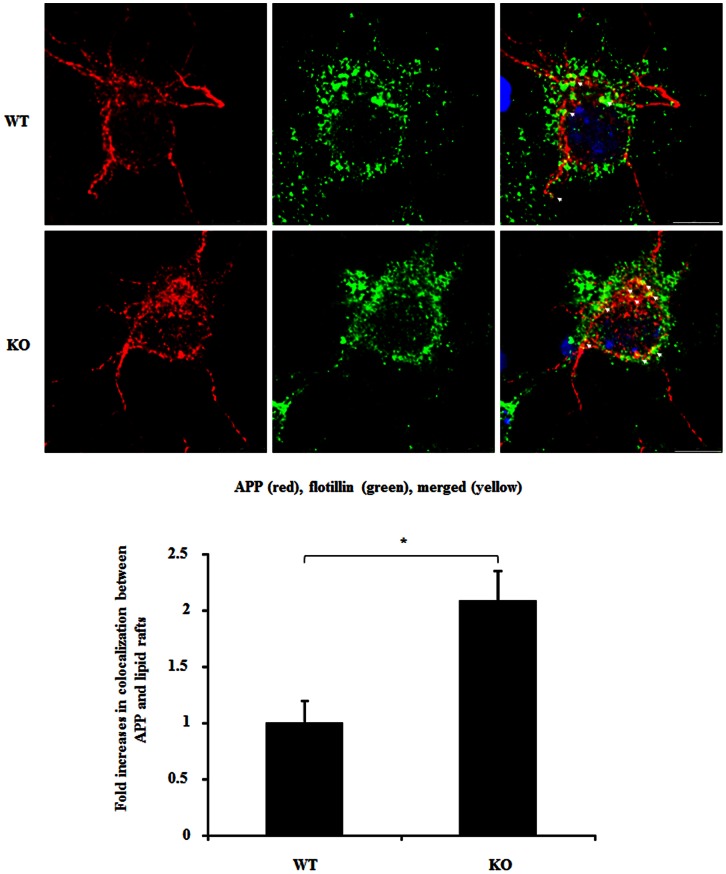
Figure 7. Lack of sortilin increases APP distribution in lipid rafts in cortical neurons.
Wild type (WT) and sortilin knockout (KO) mouse cortical neurons were immunostained for APP with mouse anti-APP-N’ (22c11) and followed by staining with Cy3 conjugated secondary antibodies (red), and lipid rafts were immunostained with anti-flotillin, followed by staining with Alexa 488 conjugated secondary antibodies (green). Cell nuclei are stained by DAPI (blue). Colocalization is analyzed by counting the number of merged APP/raft lipids (yellow), and is plotted as mean of fold increase ± SEM (n = 20 neurons). The star (*) indicates p<0.01. Scale bar 7.5 µm.

