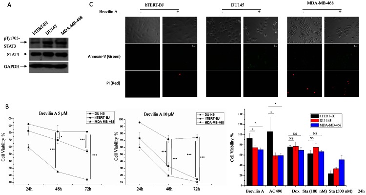
Figure 4. Brevilin A selectively inhibits cell growth of DU145 and MDA-MB-468 cells.
(A) STAT3 tyrosine 705 phosphorylation was detected in hTERT-BJ, DU145 and MDA-MB-468 cells. (B) Left and middle diagrams, hTERT-BJ, DU145 and MDA-MB-468 cells were plated in 96-well plates at a density of 8×10³ (100 µl/well in DMEM with 10% FBS). Twelve hours later, media were removed and replaced with fresh media in the presence of Brevilin A (5 µM and 10 µM) for 24 h, 48 h and 72 h, cell viability was measured by MTT assay. Right histogram, 12 hours later cells were plated, media were removed and replaced with fresh media in the presence of Brevilin A (10 µM), AG490 (100 µM), Doxorubicin (1 µM) and Staurosporine (100 nM and 500 nM) for 24 h. Dox, Doxorubicin. Sta, Staurosporine. Bars show the standard deviation (±SD) (3 independent repeats, n = 3 in each repeat). ***p<0.001, **p<0.01, *p<0.05. NS, no statistical significance. (C) Cells were plated in 24-well plates. Twelve hours later, media was removed and replaced with fresh media in the presence of 10 µM Brevilin A for 24 h. DMSO was used as control. Cells were then subjected to an Annexin-V-PI dual staining process. Same exposure program was used at each wavelength.

