FIG. 1.

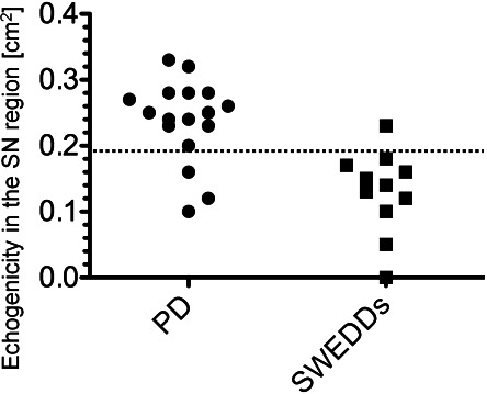
Significantly increased area of midbrain echogenicity in the PD group compared with the SWEDD group (P < 0.001) (broken line: value set at 0.20 cm2 to define hyperechogenicity).

Significantly increased area of midbrain echogenicity in the PD group compared with the SWEDD group (P < 0.001) (broken line: value set at 0.20 cm2 to define hyperechogenicity).