Figure 4.
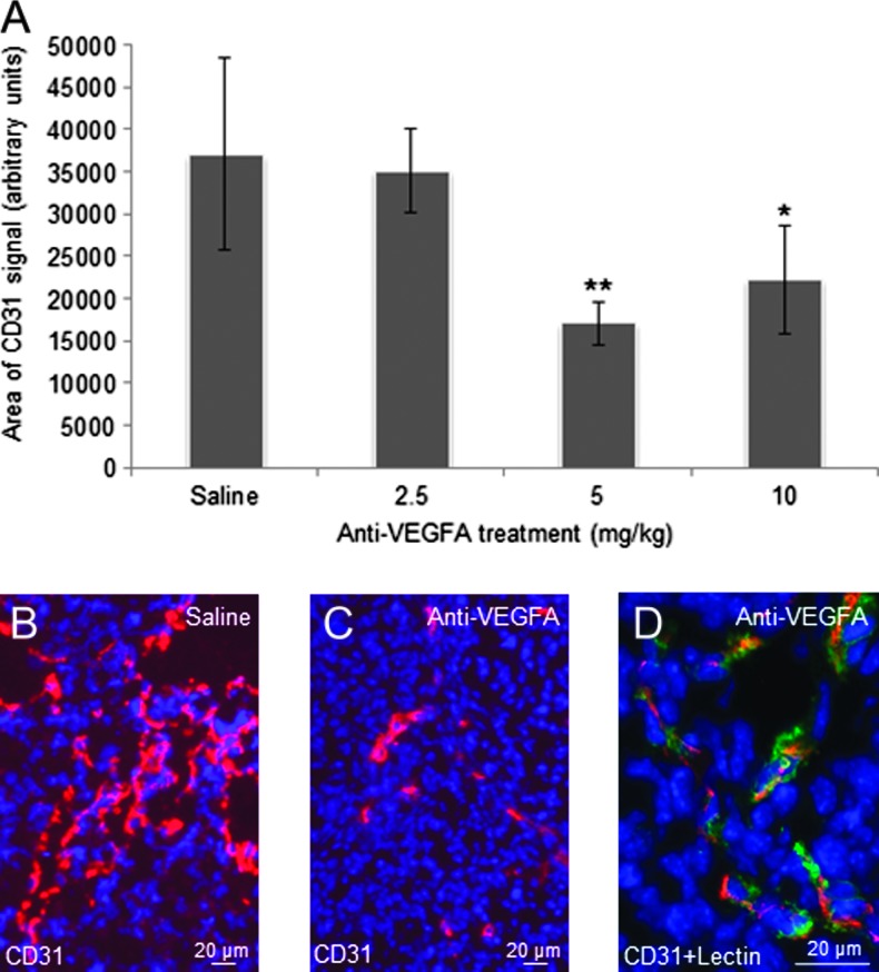
Anti-VEGFA antibody significantly reduces microvessel density in GCTs from 6-week-old PCA mice. (A) Graph depicting CD31 immunofluorescence signal strengths in GCTs from 6-weekold PCA mice that received the indicated treatments (n = 4 animals/treatment). Data are shown as means (columns) ± SEM (error bars). Significant difference from control (saline) is indicated with one (*P < .05) or two asterisks (**P < .01). (B) Representative photomicrograph (original magnification, x200) depicting CD31 fluorescent signal in the GCT of a 6-week-old PCA mouse. CD31-specific signal is red; nuclei are counterstained with DAPI (blue). (C) As per B, showing a tumor from an anti-VEGFA-treated mouse. (D) As per B, except endothelial cells were labeled with tomato plant lectin (green) in addition to CD31 immunolabeling; original magnification, x630. Overlap in lectin and CD31 signals appears yellow and confirms the specific labeling of endothelial cells by CD31.

