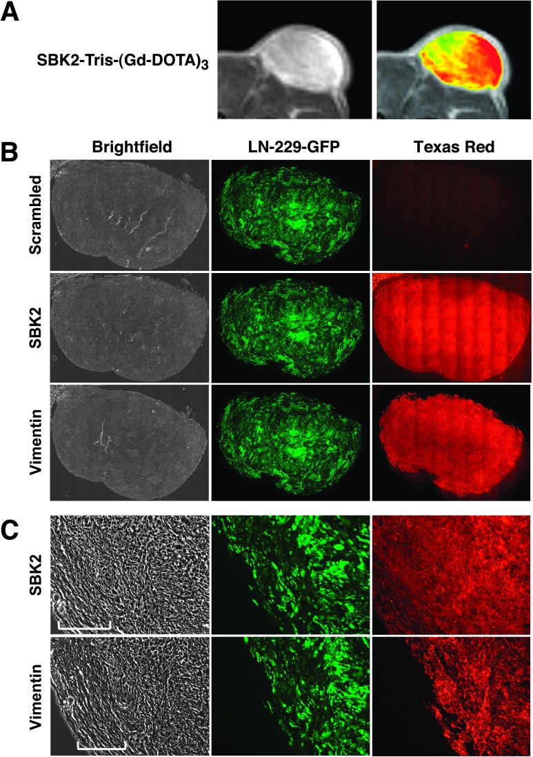
Figure 7.
Tumor labeling and histology. LN-229-GFP flank tumor MRI and histologic sections show PTPµ probe labeling of tumors. (A) Zoomed grayscale and heat map images are shown by MRI following administration of SBK2-Tris-(Gd-DOTA)3. (B) The corresponding histologic sections were labeled with SBK2-Texas Red or scrambled Texas Red probes, or anti-human vimentin antibody. Images were acquired across the entire tissue using bright-field and fluorescence optics for fluorescein (GFP) and Texas Red (probe and antibody). The resultant images were tiled and flattened to form a single composite image using Metamorph software. Anti-human vimentin antibody recognizes the human glioma tumor cells. There is co-registration of the GFP-positive and vimentin-positive cells in these tumors. (C) Zoomed images of the histologic sections are shown for each type of staining. The bracketed region in C indicates the encapsulated region surrounding the tumor.

