
Figure 1.
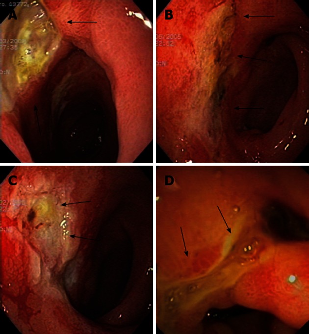
Figures of patient 5. A: A big and deep ulcer was seen in the pyloric channel and duodenal bulb at diagnosis (arrows); B: The same ulcer as in (A) in the same location seen 3 mo later, partially healed (arrows); C: Ulcer located in the duodenal bulb (arrows) with an irregular and friable mucosa after 11 mo; D: Endoscopical view of antral, pyloric and duodenal bulb deformity (arrows) seen with endoscopic ultrasound scope 78 mo after diagnosis.