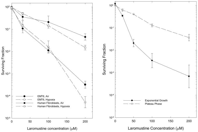
Figure 1.
Effect of hypoxia and proliferative state on the response of cells in vitro to Laromustine. Left panel: Hypoxia was induced 2 hr before treatment and continued throughout the 2 hr drug treatment. Points are means ± SEM of 4–8 independent measurements for EMT6 cells (circles) and of 3–4 measurements for human fibroblasts (squares). Surviving fractions are calculated relative to untreated control cultures; surviving fractions shown at zero dose are those for vehicle-treated control cultures subjected to all experimental manipulations. Right Panel: Survival of EMT6 cells treated with Laromustine in exponential growth or plateau phase for 2 hrs, then plated for colony formation immediately after treatment. Points are means ± SEM from 3 independent experiments.

