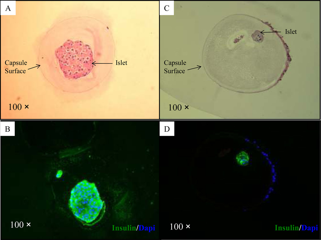
Figure 6. Immunohistochemical analysis of microbeads retrieved at day 232 after transplantation with encapsulated human islets.
(A–B): H&E staining (A) and immunofluorescent staining (B; green = insulin and blue = fluorescent DNA dye, Dapi) of a microbead retrieved from a normoglycemic mouse (mouse 1, Table 1); (C–D): H&E staining (C) and immunofluorescent staining (D) of a microbead retrieved from a hyperglycemic mouse (mouse 2, Table 1).

