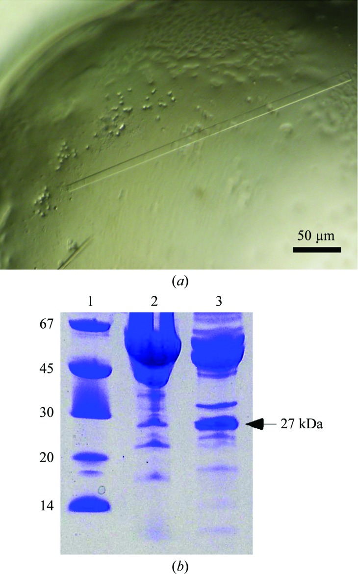
Figure 1.

(a) Triosephosphate isomerase crystals. The figure represents a portion of the crystallization drop in the PDIR crystallization screen. The typical length of the TIM crystals is 100–400 µm. (b) SDS–PAGE of PDIR (22–519) and endo-α-1,2-mannosidase crystallization samples after two-step purification. The main bands correspond to the expected molecular weights of the target proteins. The protein band at 27 kDa corresponds to triosephosphate isomerase. Lane 1 contains molecular-weight markers (labelled in kDa).
