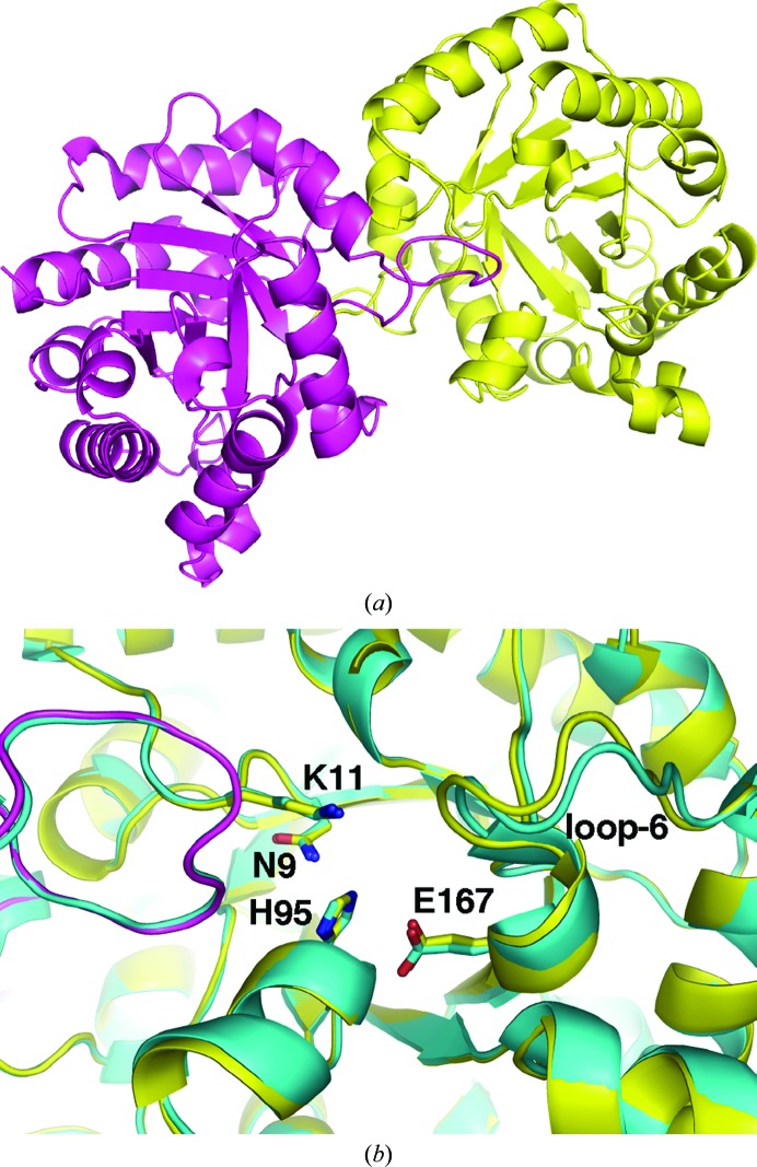
Figure 2.
Structure of E. coli triosephosphate isomerase. (a) View of the dimer comprised of TIM-barrel folds. (b) Enlarged view of the active sites of overlaid E. coli TIM structures from this work (yellow) and the previously determined structure (PDB entry 1tre; cyan). Loop 6 adopts a different conformation in the structures. Residues that are involved in the catalytic activity of TIM are shown as sticks.

