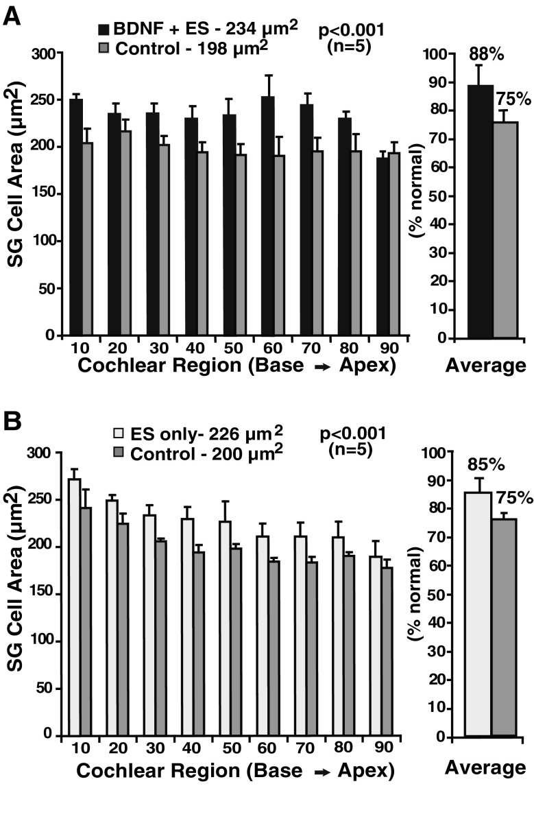
FIG. 2.
Cross-sectional areas of the somata of spiral ganglion (SG) neurons. A Data are shown in the BDNF + ES group for nine cochlear sectors representing 10 % intervals from base to apex. Cell areas throughout the cochlea are significantly larger in the BDNF + ES cochleae as compared to the contralateral deafened cochleae. After BDNF + ES, the mean SG cell area is about 235 μm2, which is significantly smaller than normal but also significantly larger than cells contralaterally, which on average measured about 200 μm2. B Data for neonatally deafened animals that received chronic ES only (no BDNF) and were selected to match the BDNF + ES group for duration of deafness and duration and type of applied ES. Again, cell areas are larger in the implanted and stimulated cochleae than contralateral, and values are quite similar to the BDNF + ES group. (Data presented are means and standard errors.)

