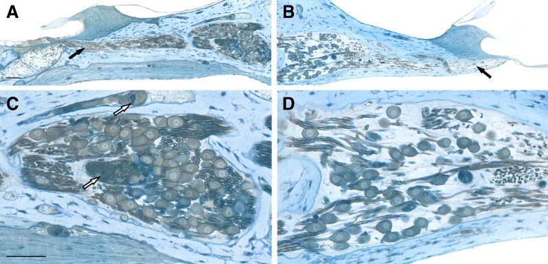
FIG. 5.
Light microscopic images of histological sections of the cochleae from a neonatally deafened animal (K313) illustrating the marked neurotrophic effects observed following combined BDNF + ES. The 20–30 % sector of the implanted left BDNF + ES cochlea (A, C) and the paired region from the contralateral deafened control cochleae (B, D) illustrate the higher density of SG cells in Rosenthal’s canal and the maintenance of a greater number of radial nerve fibers within the osseous spiral lamina (filled arrows in A, B) after BDNF infusion. In the implanted ear, the scala tympani aspect of the osseous spiral lamina (just below the radial nerve fibers) is thickened by ectopic neo-osteoneogenesis, and below this, the fibrous tissue that encapsulated the CI electrode is evident in the scala tympani (as measured for the data presented in Figure 9B). The higher magnification images in C and D illustrate the quality of preservation, staining of the tissue after osmium post-fixation and the resolution of images used for morphometric analyses in the study. The higher density and size of SG perikarya in the BDNF-treated cochlea as compared to neurons on the opposite side are evident. Open arrows in panel C indicate enlarged blood vessels after BDNF + ES as measured for data in Figure 9A. Scale bars = 50 μm.

