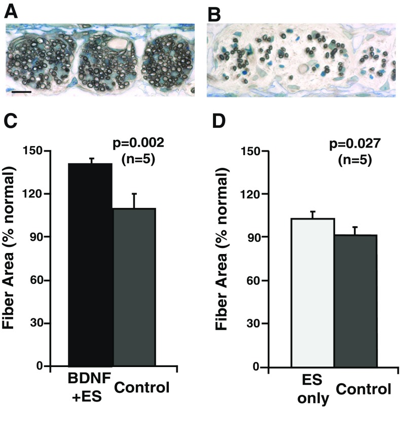
FIG. 7.
The cross-sectional areas of the radial nerve fibers were also evaluated at the same three cochlear regions where fiber counts were performed. These measurements were made in higher magnification images, as illustrated here in sections taken from the implanted cochlea (A) and contralateral deafened cochlea (B) in one combined BDNF + ES subject (K318). No systematic difference across regions was observed, and the data were averaged for all regions. C Fibers in the BDNF + ES cochleae are significantly larger than fibers in the contralateral cochleae and also markedly larger than normal. D Fiber areas in cochleae examined after chronic ES-only also are larger than fibers in the contralateral deafened cochleae, but not larger than normal.

