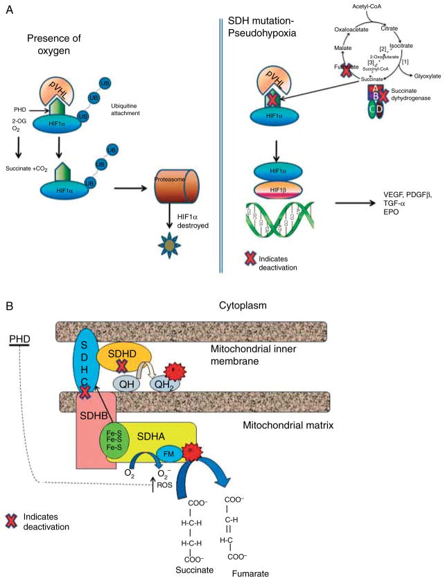
Figure 1.
(a) Mechanisms of pseudohypoxia in inherited PHEO and/or PGLs: inactivation of SDH leads to the abnormal stabilization of HIFs in normoxia that escape degradation and translocate to the nucleus, where they dimerise with HIF1β and promote transcription of genes that enhance tumorigenesis, e.g. VEGF, PDGF-β (2-OG, a-ketoglutarate; HIFs, hypoxia-inducible factors; PHD, prolyl hydroxylases; SDH, succinate dehydrogenase; VHL, Von Hippel–Lindau). (b) Oxidation of succinate to fumarate transfers the electrons through a sequence of steps from the flavin moiety in SdhA to a set of three iron–sulfur clusters in SdhB, to the ubiquinone binding site in SdhC and SdhD. When complex II is disrupted due to mutations in SdhB, SdhC, or SdhD, the electron transfer is impaired promoting superoxide generation through the autoxidation of the reduced flavin group by O2 in the matrix (FM, flavin moiety; Fe-S, iron–sulfur clusters; e−, electron transfer; QH, ubiquinone; QH2, ubiquinol).

