Figure 5.
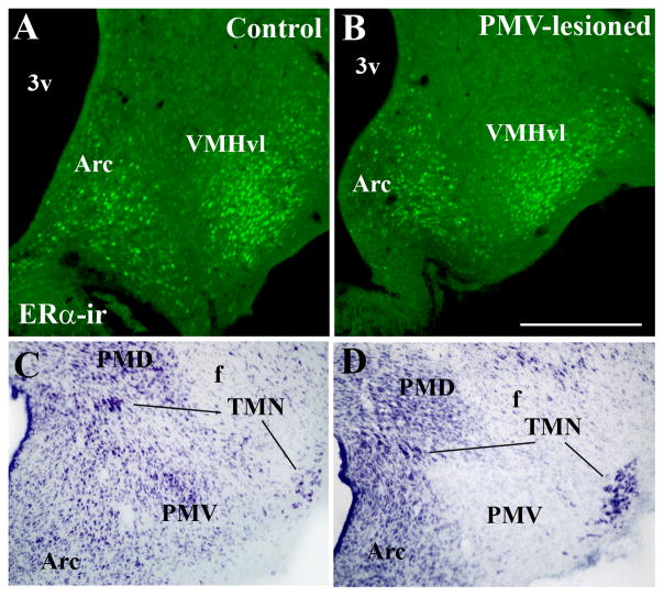
Lack of contamination of the ventromedial and the tuberomammillary nuclei by the excitotoxic lesions (NMDA injections) of the ventral premammillary nucleus (PMV). A–B, Images showing distribution of estrogen receptor α (ERα) immunoreactivity in the arcuate nucleus (Arc) and in the ventrolateral subdivision of the ventromedial nucleus of the hypothalamus (VMHvl) in a control (PMV non-lesioned, A) and in a PMV-lesioned (B) rat. Note the similarity of ERα-ir distribution in both cases, indicating lack of contamination of the VMHvl. C–D, Images showing the distribution of neurons of the tuberomammillary complex (dorsal and ventral tuberomammillary nuclei, TMN) in a control (PMV non-lesioned, C) and in a PMV-lesioned (D) rat. Note the similarity of TMN neurons (magnocellular and dark cytoplasm) distribution indicating lack of compromise of TMN neurons. Abbreviations: 3v, third ventricle; f, fornix; PMD, dorsal premammillary nucleus.

