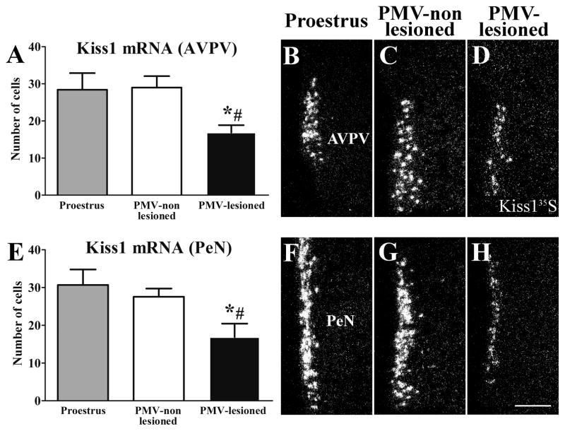
Figure 6.
Lesions of the PMV suppressed Kiss1 mRNA expression in the anteroventral periventricular nucleus (AVPV) and the periventricular nucleus (PeN) during the behavioral test (night of estrus). A, E. Bar graphs showing quantification of the number of cells expressing Kiss1 mRNA (Kiss135S) of rats perfused in the afternoon of the proestrus (n = 5), of PMV-non-lesioned rats (n = 10) and of PMV-lesioned rats (n = 8) in the AVPV (A) and the PeN (E). PMV-lesioned rats showed a decreased number of Kiss1 cells in the AVPV and the PeN after the sexual behavior test compared to proestrus and PMV-non-lesioned rats. B–D, F–H. Darkfield photomicrographs showing the expression of Kiss1 mRNA in a proestrus (B, F), a PMV-non-lesioned (C, G) and a PMV-lesioned (D, H) rat in the AVPV (B–D) and the PeN (F–H). Data in bar graphs are expressed as mean ± SEM. *, statistically different (P < 0.05) from the proestrus group. #, statistically different (P < 0.05) from the PMV-non-lesioned group. For statistical analysis, we used the one-way ANOVA followed by the pairwise Newman-Keuls test. Scale bar = 200 μm.

