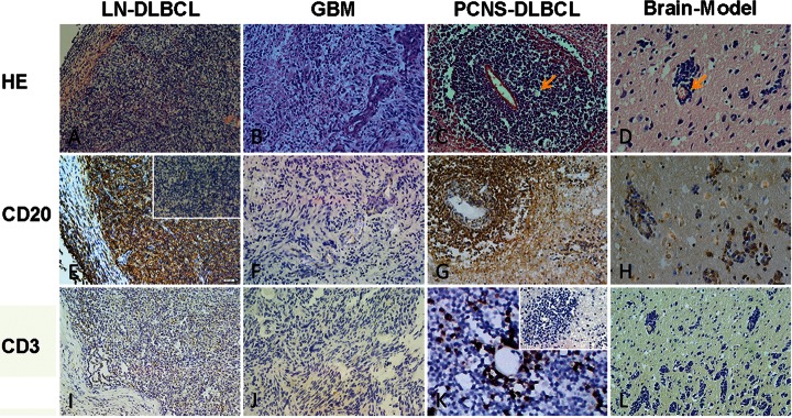
Fig. 1.

HE, CD20, and CD3 staining of the lymph node–diffuse large B cell lymphoma (LN-DLBCL), glioblastoma (GBM), primary central nervous system–diffuse large B cell lymphoma (PCNS-DLBCL), and Brain-Model. A, B, C, and D represent HE staining of LN-DLBCL, GBM, PCNS-DLBCL, and Brain-Model. PCNS-DLBCL and Brain-Model display clear APVT (yellow arrow), whereas both LN-DLBCL and GBM do not have APVT. E, F, G, H and I, J, K, L represent CD20 and CD3 staining of LN-DLBCL, GBM, PCNS-DLBCL, and Brain-Model, respectively. The cells with brown coloration are positive. In PCNS-DLBCL, CD3-positve T cells accumulated around the perivascular area interspersed within tumor cells (RPVI). The inserted figures are negative controls (magnification ×200).
