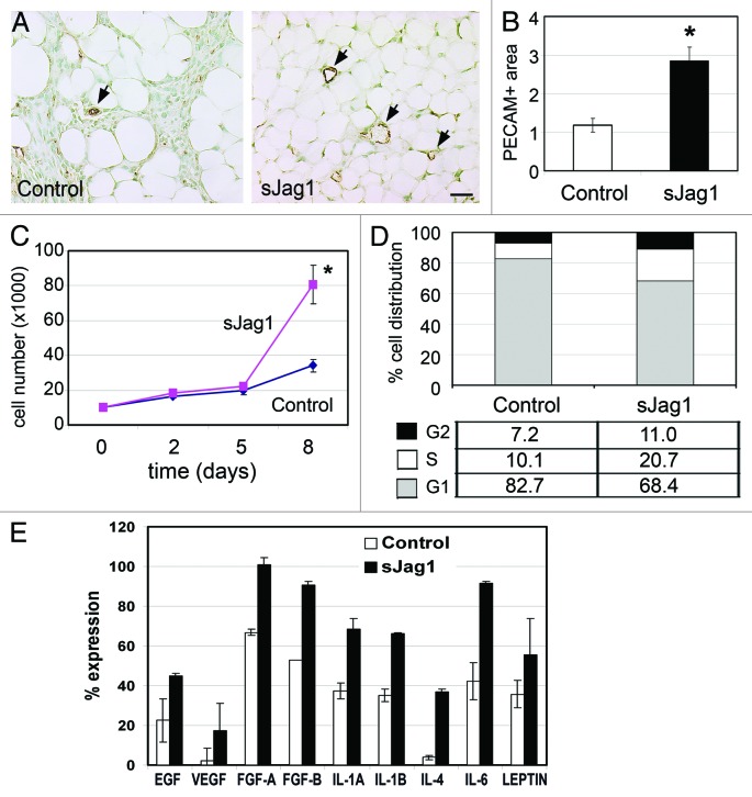
Figure 5. sJag1 cells promote angiogenesis and endothelial cell proliferation. (A) Representative fat pad sections from subcutaneous injections stained with PECAM to determine vessel density (bar = 50 μm). (B) The graph shows quantification of the vessel area (n = 6 tumors; 20 pictures each; graphed are means ± SD and * indicates p < 0.05). In vitro effect of secreted sJag1 on endothelial cell. (C) Growth curve analysis of HUVEC cells treated with control or sJag1 conditioned media (graphed are means ± SD and * indicates p < 0.05). (D) Cell cycle analysis showing distribution of endothelial cells at G1, S and G2 phase cultured in cell-free conditioned media from control or sJag1 3T3- L1 cells. (E) ELISA based immuno-analysis of pro-angiogenic cytokines secreted from control and sJag1 cells. The graph shows quantification of the immunoblot based on integrated densities (graphed are means ± SD).

An official website of the United States government
Here's how you know
Official websites use .gov
A
.gov website belongs to an official
government organization in the United States.
Secure .gov websites use HTTPS
A lock (
) or https:// means you've safely
connected to the .gov website. Share sensitive
information only on official, secure websites.
