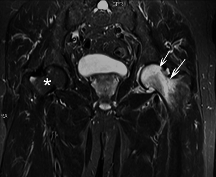
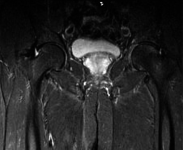
Figure 2.
A) Initial coronal fluid–sensitive (STIR) MR image obtained 3 months after initial onset of pain reveals hyper-intense bone marrow signal within the left femoral head and neck (arrows) consistent with the bone marrow edema associated with transient osteoporosis. A joint effusion is also present. The right femoral head and neck exhibit normal bone marrow signal. (*)
B) Follow up coronal fluid–sensitive (STIR) MR image obtained 3 months after the initial MR study reveals complete resolution of the abnormal bone marrow signal within the left femoral head and neck as well as resolution of the initial joint effusion. The right femoral head and neck exhibit normal bone marrow signal.


