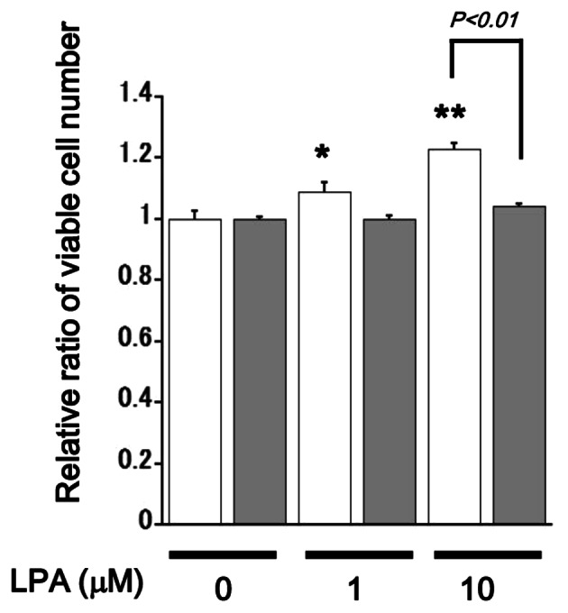
Figure 5.

Proliferation assay of AdvLPA4G-infected SQ-20B cells. Cells were infected with 100 MOI of AdvLPA4G and were then incubated for 24 h with (gray bars) or without (open bars) 100 ng/ml of Dox in SFM. WST-1 assay was performed 48 h after the LPA stimulation (concentrations are indicated). Data are shown as the mean ± SEM (n=6). *P<0.05; **P<0.01 against the control unstimulated cells. P-values are indicated; ns, not significant.
