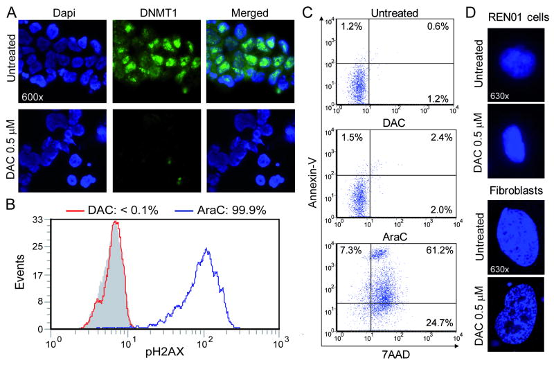
Figure 1. Decitabine (DAC) 0.5 μM depletes DNMT1 in Ren-01 cells without causing significant DNA damage or apoptosis. A) DNMT1 depletion in Ren-01 cells treated with decitabine 0.5 μM.
Ren-01 cells (low passage number RCC cells) at <40% confluence were treated with decitabine 0.5 μM. DNMT1 was quantified 48 hours later by immuno-fluorescence (green dots). DAPI was used to stain nuclei (blue stain). B) This concentration did not produce measurable DNA damage in Ren-01 cells. 24h after DAC or AraC exposure DNA damage was measured by flow-cytometric assessment for phosphorylation of histone H2AX. Equimolar levels of AraC used as positive control. Grey histogram = isotype control. C) Decitabine 0.5 μM did not produce early apoptosis in Ren-01 cells. 24h after addition of DAC or AraC 0.5 μM, apoptosis was measured by flow-cytometric assessment for Annexin/7AAD staining of exposed phosphatidyl-serine. D) Decitabine produced chromatin changes associated with senescence in normal fibroblasts but not in Ren-01 cells. Normal human fibroblasts, but not Ren-01 cells, treated with decitabine undergo clumping changes in chromatin associated with senescence 25.

