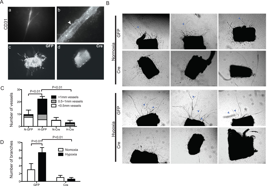
Fig. 1.
Loss of ARNT inhibits hypoxia enhanced vessel outgrowth and branching in aortic-ring explants. (A) Magnification of vessel outgrowth from rings stained for Rhodamine-CD31 (a, b). Note the vacuolated vessels in b (Arrowhead). Fluorescent micrographs of rings dissected from aortas of ArntloxP/loxP adult mice were infected with Ade -GFP (c) or -Cre/GFP (d). (B) Phase-contrast micrographs of representative rings infected with Ade -GFP or -Cre/GFP after culturing for 10 days under normoxic or hypoxic (2.5% O2) conditions. Arrows indicate vessel branches. Quantification of vascular outgrowth assessed by the number of vessels, length of vessels (C) and branches (D). Data shown are the mean ± SEM, n=10 samples. Statistical analysis was performed using an unpaired Student’s t-test.

