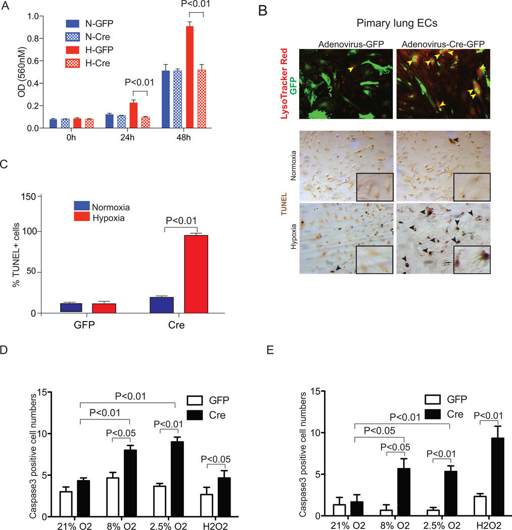
Figure 4.
Arnt-null endothelial cells display proliferation and survival defects. ArntloxP/loxP primary lung and heart endothelial cells (ECs) were infected with Adenovirus -GFP or -Cre/GFP. (A) BrdU incorporation to assess lung EC proliferation under normoxic or hypoxic (2.5% O2) conditions. (B) Upper panels, Lysotracker Red (LTR) staining detecting cellular death in primary lung ECs. Arrows, LTR+/Cre+ cells. Middle/lower panels, TUNEL staining (brown) and quantification (C) of primary lung ECs cultures 24 hours following serum withdrawal in 2.5% or 21% O2 conditions. High magnification images are shown in the bottom right corner of each panel. (D,E) Quantification of Caspase-3 from primary lung (D) and heart (E) endothelial cells in response to 21, 8, or 2.5% O2for 24 hours or 300uM H2O2 for 2 hours.

