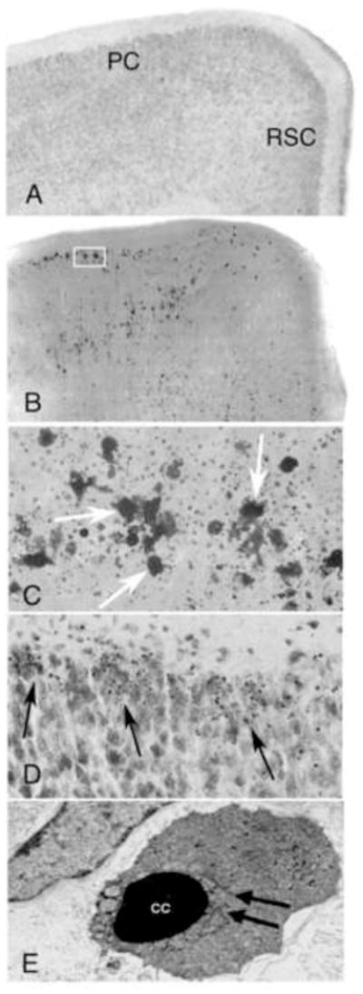
Figure 1.

Coronal hemi-sections from the brain of a 7-day-old mouse treated with saline (A) and an animal treated with lead acetate 24 hours previously (B) stained by the De Olmos silver method. Degenerating neurons (dark areas) are abundantly present throughout, particularly in the parietal and retrosplenial cortices (PC and RSC, respectively). The box indicates the region illustrated in the next panel (C) at a higher magnification. Neurons (arrows) are in an advanced stage of degeneration. Their amorphous shape and sourrounding debris makes it difficult to determine their neuronal type. Image (D) demonstrates a Nissl stained section of parietal cortex in a lead treated animal at 24 hours. Abundant dense chromatin spheres (i.e. apoptotic bodies) are present (arrows). Image (E) is an electron micrograph of a degenerating neuron taken from a section of the parietal cortex. Neuron is condensed and has clumped chromatin (CC) with fragmentation of the nuclear membrane (arrows), verifying that the degenerative process is apoptotic.
