
Figure 2.
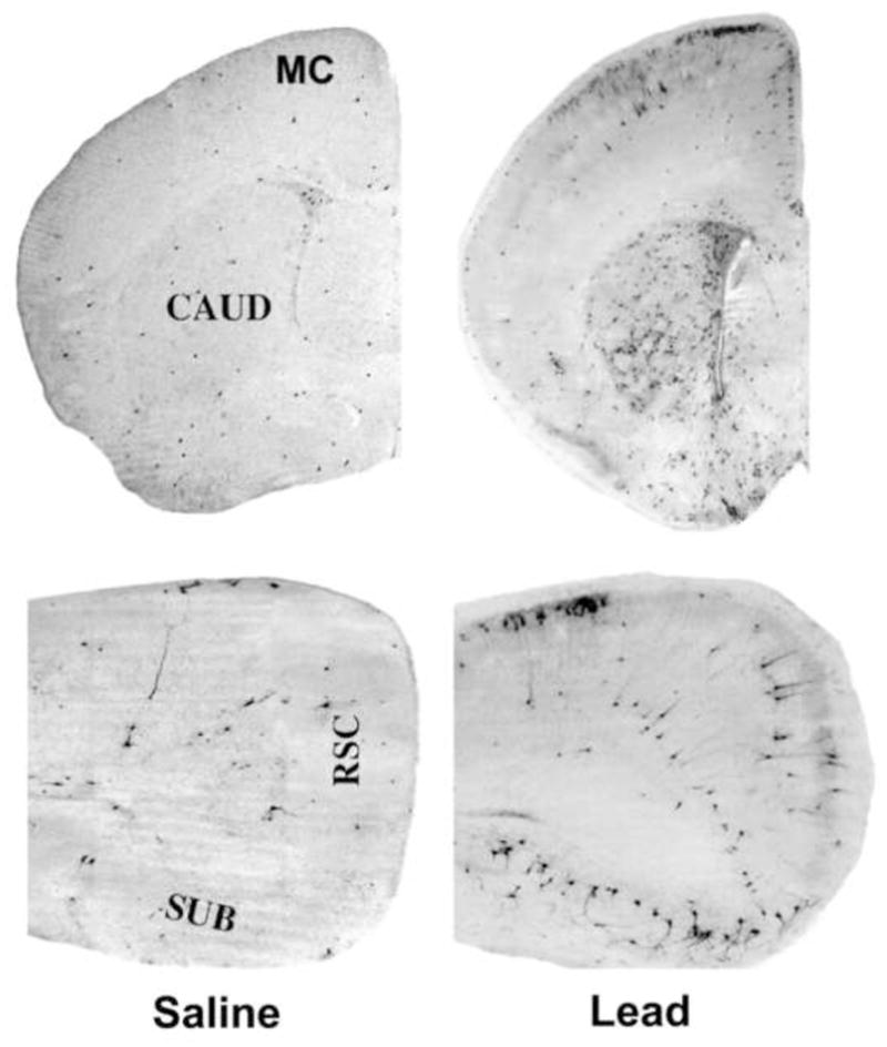
Coronal hemi-sections of 7-day-old mice treated with saline or lead acetate. Sections were stained immunohistochemically with antibodies to activated caspase-3. The saline control brain (left) shows a pattern of caspase-3 activation that occurs normally in the 7-day-old mouse brain and is attributable to physiological cell death. Neurons displaying activated caspase-3 are prevalent in several brain regions of the lead-exposed animal. Notable areas of involvement include the anterior motor cortex (MC), caudate nucleus (CAUD), subiculum (SUB), and retrosplenial cortex (RSC).