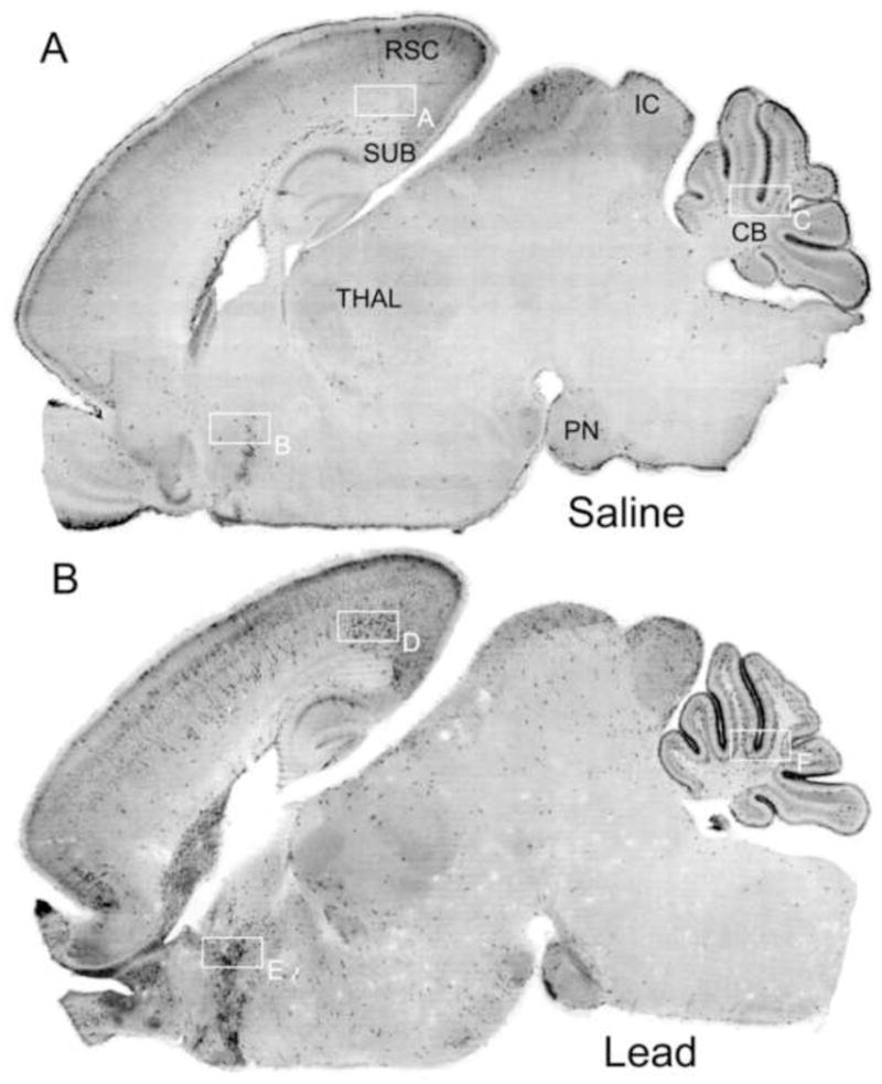
Figure 3.

Sagittal sections of 7-day-old mice treated with saline or lead acetate immunostained with antibodies to activated caspase-3. Image (A) is a saline control brain showing a pattern of caspase-3 activation that occurs normally in the 7-day-old mouse brain and is attributable to physiological cell death. Image (B) is the brain from a lead-treated animal. Numerous neurons displaying activated caspase-3 are prevalent in several brain regions including: the subiculum (SUB), retrosplenial cortex (RSC), and diffuse layers of the superficial and deep cortices; the inferior colliculis (IC), pontine nucleus (PN), and cerebellum (CB). High power photomicrographs of panels A through F are shown in figure 4.
