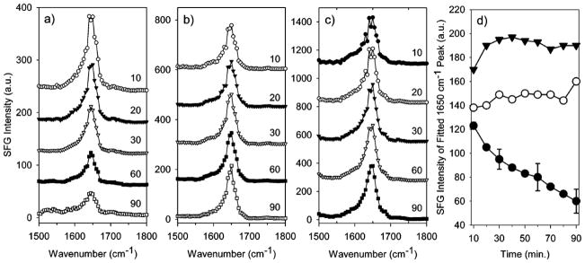
Figure 18.

SFG spectra collected in the amide I range of fibrinogen adsorbed to (a) PEU, (b) SPCU, and (c) PFP in PBS buffer at different time (in min). Time dependent SFG signal of α-Helix (d) from fitting SFG spectra for fibrinogen adsorbed to PEU (closed circles), SPCU (open circules), and PFP (closed triangles). Representative error is shown for the fibrinogen/PEU sample. Reprinted with permission from ref. 200. (2005 American Chemical Society)
