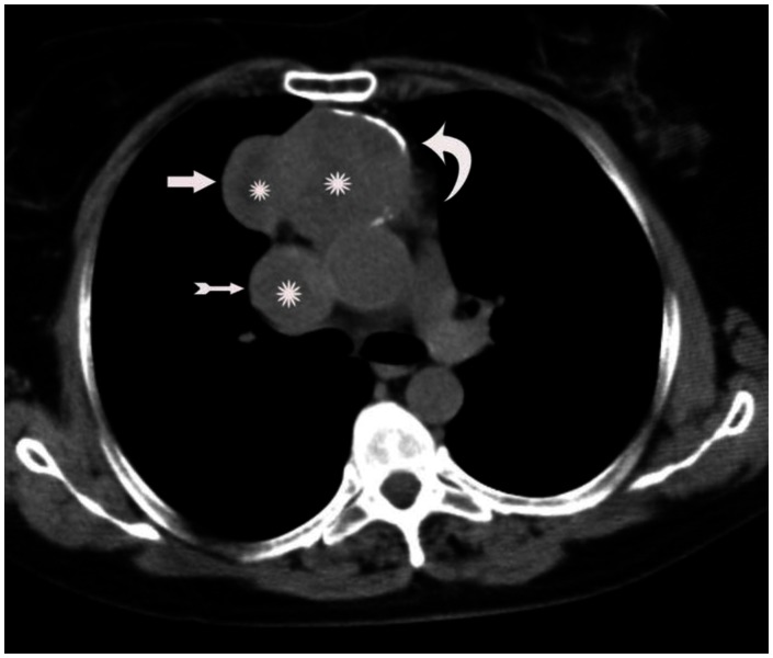
Figure 1.
A 53-year-old woman with invasive thymoma with extensive intraluminal tumor thrombus. Axial plane non-contrast-enhanced CT shows anterior mediastinal mass with lobulated contours (thick arrow) and rim calcification (curved arrow). Note the enlarged superior vena cava (thin arrow). The mass and the thrombus are heterogeneous and central parts are more hypodense consistent with necrosis (stars).(Protocol: 64 detector scanner, 180 mAs, 120 kV, slice thickness: 5 mm.)

