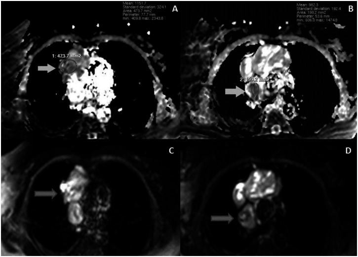
Figure 6.
A 53-year-old woman with invasive thymoma with extensive intraluminal tumor thrombus. ADC maps (A, B) and DWI (C, D) of the mass and the thrombus at same level. The mass (A, arrow) and the thrombus (B, arrow) show significant low intensities on ADC maps with low ADC values, compatible with the malignant character. (Protocol: 3 Tesla MRI, TR:1763, TE:69)

