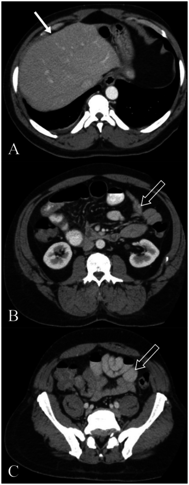
Figure 3.
A 49-year-old male with resolved cocaine-induced mesenteric ischemia. Axial CT images (Toshiba Aquilion 32 slice, 121 mAs, 120 kVp, 3 mm axial slices, 120 cc of Optiray 350 contrast in portal venous phase, 900 cc of Readi-CAT given 2 hours prior to image acquisition) with intravenous and positive oral contrast 24 hours after initial presentation. Images demonstrate normal appearance of small bowel (3b and 3c open straight arrow) and resolution of the perihepatic free fluid (3a closed straight arrow). No abnormal findings were noted.

