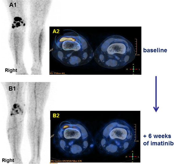
Figure 3.

[18F]fluorodeoxyglucose (FDG) PET/CT tumor assessment of Patient 2. Baseline PET/CT maximum projection image (MIP) (panel A1) showed abnormal FDG focal uptakes in the right knee PVN with a SUVmax of 14.7 g/ml, as detailed by fused PET/CT transaxial slice (panel A2). After 6 weeks of treatment, PET/CT MIP (panel B1) and fused PET/CT transaxial slice (panel B2) showed a marked decrease of tumor FDG uptake (SUVmax 4.1 g/ml, i.e. 72% reduction).
