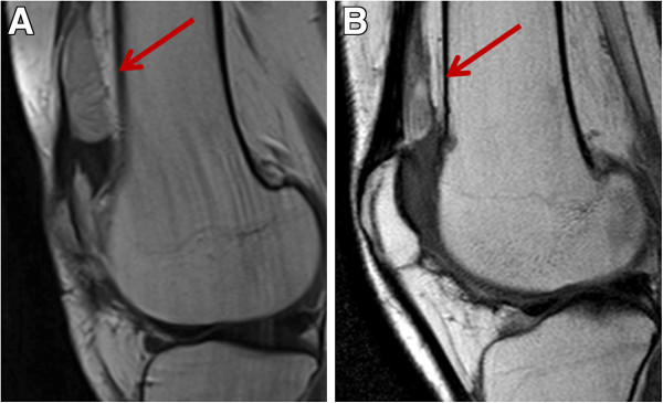
Figure 4.

Magnetic resonance imaging (MRI) tumor assessment of Patient 2. Sagittal contrast enhanced (ce) TSE T1 weighted (w) MRI images at baseline showed a lesion located behind the rotula (panel A). After 2 months of imatinib a decrease in tumor size was detected (panel B).
