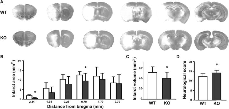
Figure 1.
β2AR knockout reduced infarct size and improved neurological deficits after transient focal ischemia. (A) Histology of cresyl violet stained brain sections was assessed at 2.34 mm, 1.34 mm, and 0.26 mm rostral, and −0.70 mm, −1.70 mm, and −2.70 mm caudal from bregma. Representative sections from brains of wild type (WT) (upper row) and knockout (KO) (lower row) mice are shown. Pale areas indicate injured tissue. (B) Analysis of infarct area at each of the 6 coronal levels in WT versus KO mice after 24 h reperfusion. (C) Total infarct volume was decreased by 22.3% in KO mice compared with WT littermates (39.7 ± 10.7 mm3 vs 51.0 ± 11.4 mm3, P = 0.034). (D) Neurological deficit score was improved in KO mice compared with WT at 24 h reperfusion (12.9 ± 1.4 in WT versus 14.7 ± 1.5 in KO, P = 0.012; n = 10 per group). *Indicates significant difference from WT. Filled bars indicate KO, open bars WT.

