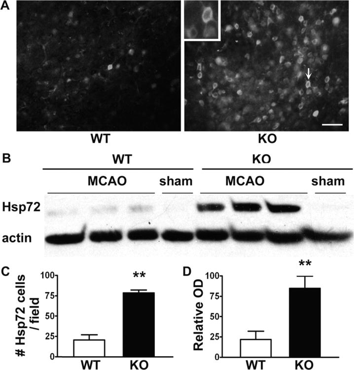
Figure 3.
Increased heat shock protein (Hsp) Hsp72 expression in mice lacking β2AR after 24 h ischemia. (A) Brain sections immunostained for Hsp72. (C) Following middle cerebral artery occlusion (MCAO) the number of Hsp72 positive cells in the penumbra increased significantly in knockout (KO) mice compared with wild type (WT) mice (21 ± 7 vs 78 ± 5, P < 0.001). (B, D) Western blots of cytosolic fractions show that Hsp72 is increased in the ischemic hemisphere of KO mice compared with WT (22.0% ± 9.0% vs 84.8% ± 13.2%, P < 0.001). Actin band intensity was used to normalize the Hsp72 band intensity. (n = 6 per group, scale bar = 40 μm).

