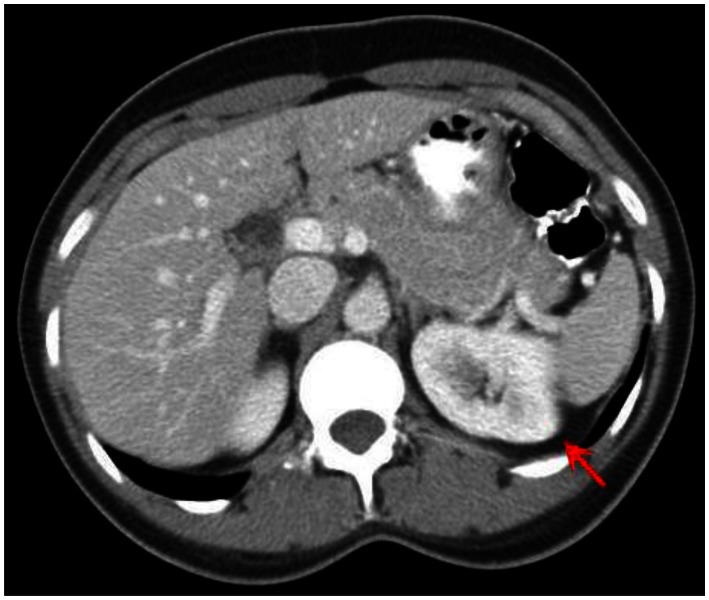
Figure 10.
53 year old female with biopsy proven renal sarcoid. Contrasted CT axial image demonstrates an ill defined cortical left renal mass (red arrow), with enhancement features similar to that of the normal kidney, but around an encapsulated lesion. CT examination of the chest and abdomen with intravenous contrast (Omnipaque 300). (CT scanner: LightSpeed 16; Mag: 1.7x; 120 kV; 100–300 mA; 5.0 mm Tilt: 0.0; ET: 0.8 s; GP: 0.6 s; TS: 17.50 mm/s; W: 400. L: 40; 512 × 512 Matrix; DFOV: 34.5 × 34.5 cm)

