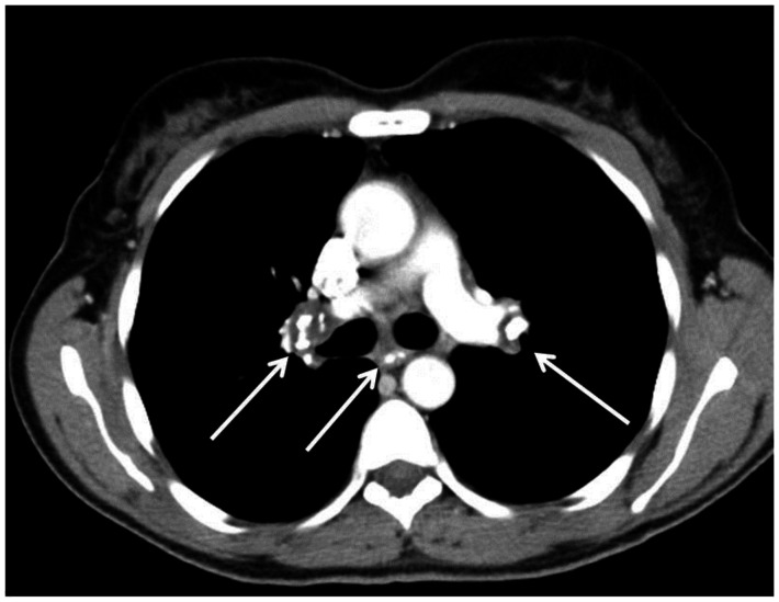
Figure 11.
53 year old female with an calcified mediastinal and hilar lymphadenopathy, known to be secondary to sarcoidosis. Contrasted axial CT images (soft tissue windows) demonstrate the calcified subcarinal and bilateral hilar lymph nodes. . CT examination of the chest and abdomen with intravenous contrast (Omnipaque 300). (CT scanner: LightSpeed 16; Mag: 1.7x; 120 kV; 100–300 mA; 5.0 mm Tilt: 0.0; ET: 0.8 s; GP: 0.6 s; TS: 17.50 mm/s; W: 400. L: 40; 512 × 512 Matrix; DFOV: 34.5 × 34.5 cm)

