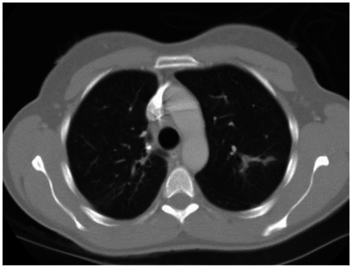
Figure 12.
53 year old female with a known history of sarcoidosis. Contrasted axial CT images (lung windows) demonstrate peribronchovascular nodularity. CT examination of the chest and abdomen with intravenous contrast (Omnipaque 300). (CT scanner: LightSpeed 16; Mag: 1.7x; 120 kV; 100–300 mA; 5.0 mm Tilt: 0.0; ET: 0.8 s; GP: 0.6 s; TS: 17.50 mm/s; W: 400. L: 40; 512 × 512 Matrix; DFOV: 34.5 × 34.5 cm)

