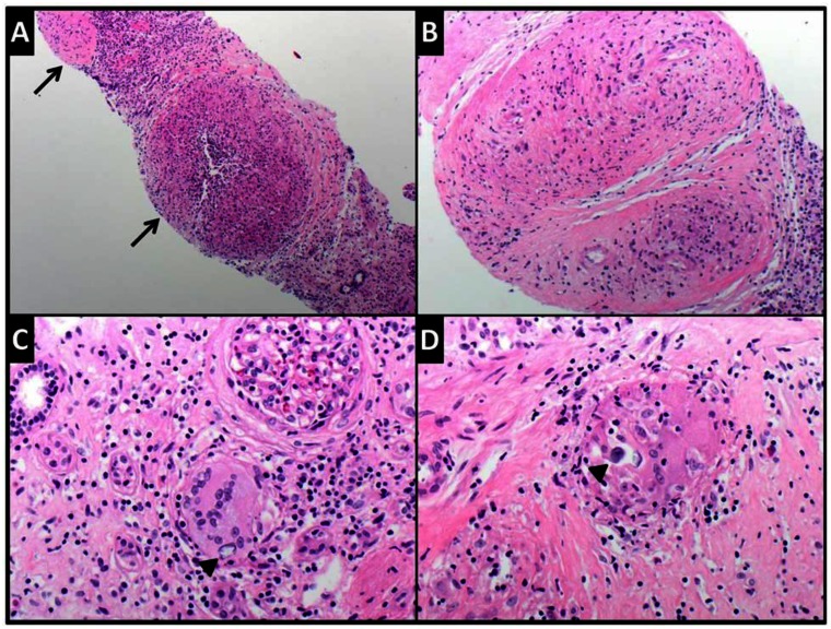
Figure 13.
Composite photomicrograph showing the microscopic findings in the renal biopsy from this case. Low (A) and intermediate (B) power views showing multiple nonnecrotizing granulomata (arrows) in renal parenchyma. They are composed of epithelioid histiocytes, including some giant forms. Giant cells with concentric calcifications consistent with Schaumann bodies (arrow heads) are shown in C and D (Hematoxylin and eosin stained-sections, original magnifications X40, X100, X200 and X400 respectively).

