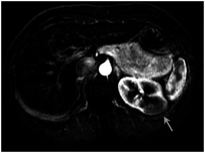
Figure 8.
53 year old female with biopsy proven renal sarcoid. Subtracted postcontrast T1 image demonstrates an ill defined exophitic left renal mass (white arrow), with mild enhancement to a lesser degree than the surrounding normal renal parenchyma. (GE Signa HDx 1.5T T1 weighted LAVA fat saturated axial sequence, TR 4, TE 2, Venous-arterial postcontrast subtraction 12cc Omniscan injection)

