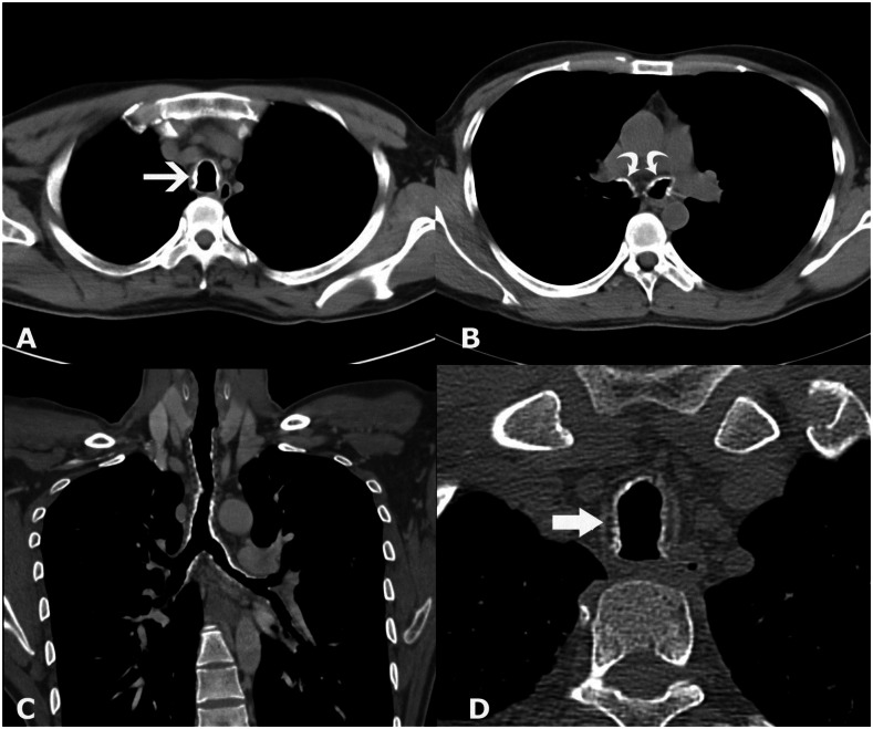
Figure 3.
28-year-old male with Tracheobronchopathia Osteochondroplastica presenting with expectoration, breathlessness and wheezing. (A) Non-contrast axial CT section shows presence of nodular wall calcification of inner tracheal lining (white arrow) with sparing of posterior wall. (B) Lower sections reveal nodular calcification in main bronchi (arrows). (C) Coronal reconstructed image reveals the extent of involvement of tracheobronchial tree. Lower two third trachea and main bronchi are affected with relatively spared upper third of trachea. (D) High resolution zoomed image shows location of calcification internal to cartilage (arrow). (128-channel MDCT scanner SOMATOM Definition AS, Siemens Medical Solutions, 120 kV, automatic mAs, detector collimation 128 × 0.6 mm, window width/level of 360/60 HU, 0.6 mm slice thickness for high resolution image and 3 mm for other images).

