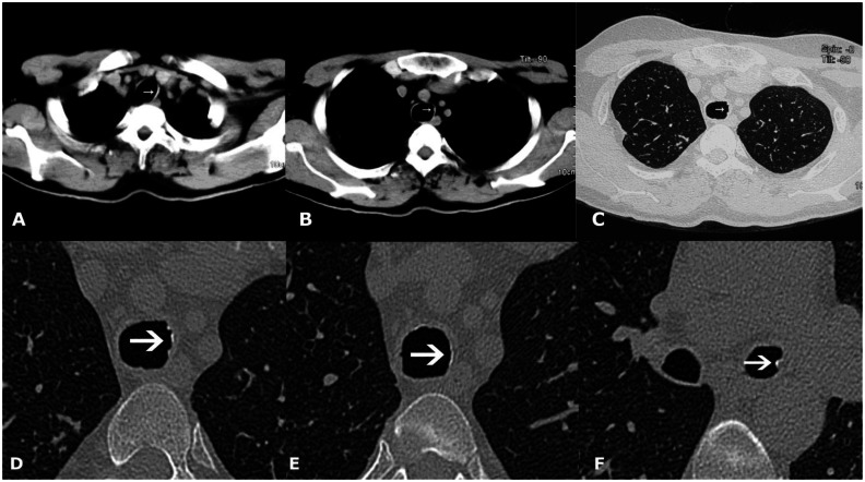
Figure 9.
42-year-old male with Tracheobronchopathia Osteochondroplastica presenting with hemoptysis. (A, B) Non-contrast axial soft tissue window CT images reveal faint calcification on the left lateral wall of trachea (white arrow). (C) High resolution lung window CT image reveals subtle nodularity of inner tracheal surface (arrow). (D, E, F) Magnified high resolution images of trachea and left bronchi showing nodular calcification (arrows). (128-MDCT scanner SOMATOM Definition AS, Siemens Medical Solutions, 120 kV, automatic mAs, detector collimation 128 × 0.6 mm, 3 mm reformation and window width/level of 360/60 HU for soft tissue images, 0.6 mm slice thickness and width/level of 1600/−600 HU for lung window image, 0.6 mm magnified high resolution images).

