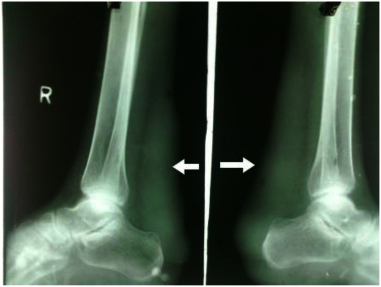
Figure 1.
30 year old male with bilateral Achilles tendon xanthomas. A plain radiograph of bilateral ankles (45 kV and 15 mAs, without grid, film-screen cassette) showing soft tissue radiodense opacity in the region of the Achilles tendon depicted with solid arrows. The visualized bones and joints in the x-ray appear normal. There is no evidence of any bony erosion seen in the calcaneum.

