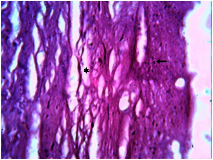
Figure 11.
30 year old male with cerebrotendinous xanthomatosis. H-E stained section of the biopsy specimen (magnification 50x) from the Achilles tendon shows degenerated fibrocollagenous tissue marked with an asterisk interspersed with adipose cells and foam cells shown with a solid arrow. The presence of Touton giant cells is a characteristic finding.

