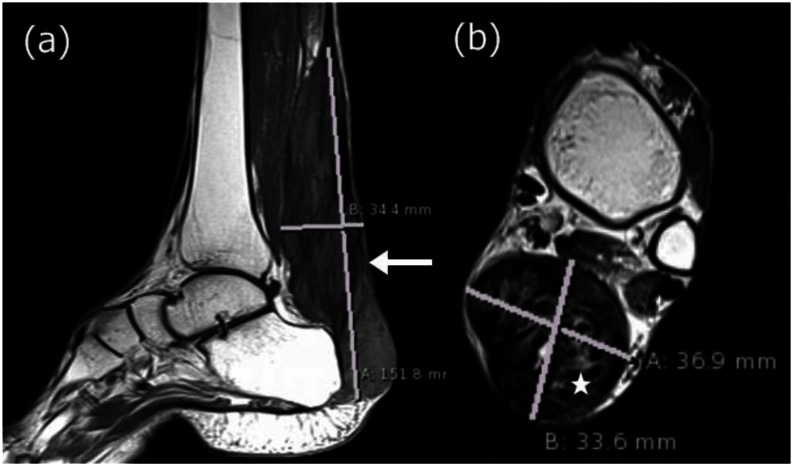
Figure 5.
30 year old male with bilateral Achilles tendon xanthomas. (a) Saggital T1W MRI (TR/TE - 400/10 ms, 3 mm slice thickness, 320×320 matrix) shows diffuse low intensity infiltration of the Achilles tendon which appears enlarged and is marked with a solid arrow. (b) Axial T2W MRI (TR/TE - 4380/84 ms, 4 mm slice thickness, 312×384 matrix) shows diffuse low intensity infiltration of the Achilles tendon with a few isointense areas marked with an asterisk. The xanthoma measures 15 cm in length, 3.4 cm in anteroposterior diameter and 3.7 cm in transverse dimension. The overall lesion is well defined with smooth margins and no infiltration into the adjacent soft tissue.

