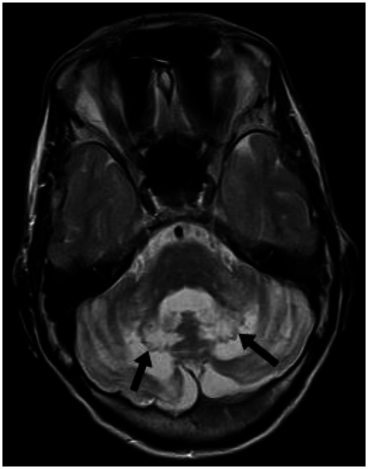
Figure 6.
30 year old male with cerebrotendinous xanthomatosis. Axial T2W MRI (TR/TE - 3800/91 ms, 5 mm slice thickness, 384×288 matrix) of the brain shows bilateral hyperintense lesions involving the dentate nuclei and the deep cerebellar white matter shown with arrows. The cerebellar cortex and the vermis appear normal in morphology and signal intensity. The brain stem appears normal in signal intensity.

