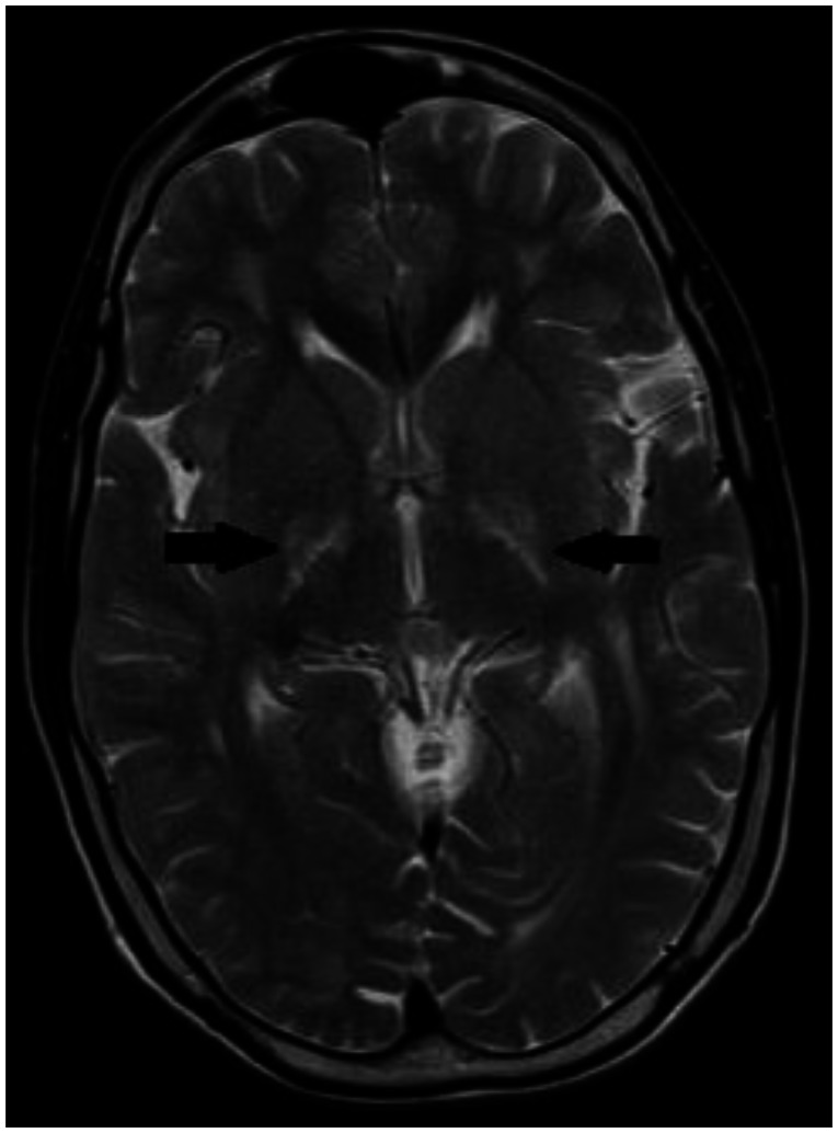
Figure 8.
30 year old male with cerebrotendinous xanthomatosis. Axial T2W MRI (TR/TE - 3800/91 ms, 5 mm slice thickness, 384×288 matrix) of the brain shows hyperintense lesions in the posterior limb of internal capsule. The head of caudate nucleus and putamen do not show any signal abnormality. Bilateral thalami appear normal. Both the lateral ventricles appear normal.

